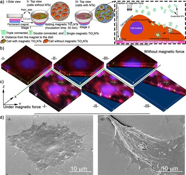
Figure 3.
(a) Schematic of enhanced cellular uptake of magnetic TiO2NTs under the influence of a gradient magnetic field. The cells were incubated with FITC-conjugated magnetic TiO2NTs for 30 min and then washed thoroughly to remove unbound nanocarriers. The overall distance between the permanent magnet and the cell surface was ≈2.7 mm. Representative micrographs of FITC-conjugated magnetic TiO2NTs without (b) and with (c) application of a gradient magnetic field imaged by progressive Z-stack confocal microscopy (ImageJ processed 3D volume micrographs). Roman numbers, indicate images at different XY distances, illustrate internalized nanocarirers (green). Cell nuclei and actin were stained with DRAQ5TM (shown in blue) and phalloidin-TRITC (red) respectively, and FITC-conjugated magnetic TiO2NTs are shown in green. (d) Representative SEM micrographs of internalizing magnetic TiO2NTs without (I) and with (II) magnetic force.

