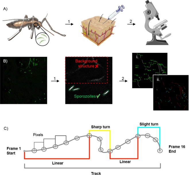
Figure 1.
Schematic of experimental setup and SMOOThuman skin analysis. (A) Schematic of an Anopheles mosquito as the host of Plasmodium sporozoites within its salivary glands. Isolated sporozoites, Pf WT or Pf RA, were injected into human skin (1). The skin samples containing sporozoites were filmed using a confocal microscope (2) (Images of needle and microscope were adapted from image copyright https://smart.servier.com, Creative Commons Attribution 3.0 Unported License, https://creativecommons.org/licenses/by/3.0/). (B) Raw confocal video images were uploaded into SMOOThuman skin. Per video frame individual sporozoites were semi-automatically segmented (1). Segmented sporozoites in consecutive frames were stitched to generate tracks. Generated tracks have a unique sporozoite ID (i) in order to extract measured parameters (for example movement pattern (ii)) per sporozoite over time (2). (C) Sporozoite tracks are divided into segments based on the underlying movement pattern.

