Fig. 5.
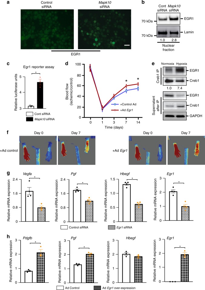
Egr1 enhances the blood flow recovery after hindlimb ischemia. a, b Neuro-2a cells were treated for 48–72 h with scramble or Mapk10 siRNA and exposed to hypoxia for 30 min. Then immunostaining was performed with an Egr1 antibody (a) and lysate was prepared after nuclear fraction isolation and immunoblotting was performed with Egr1 and control antibodies (b). c A Luciferase reporter assay for the Egr1 activity was performed on N2a cells treated with either control siRNA or Mapk10 siRNA (48 h) and 90 min of hypoxia (n = 3 in each group). d Control Ad-GFP and Ad-Egr1 were injected (single dose of 2 × 10⁸ c.f.u.) into the gastrocnemius muscle of WT mice. Three days following the injection femoral artery ligation was performed and blood flow measurements by laser speckle contrast imaging (Control Ad, n = 6; Ad-Egr1, n = 7) were obtained. e Neuro-2a cells were treated with either normoxia or hypoxia followed by immunoprecipitation (IP) with a Creb1 antibody and immunoblotting with antibodies for Egr1 and Creb1. Supernatants were examined after IP by probing with antibodies as indicated. f Representative images of hindlimb blood flow of WT mice 7 days post femoral artery ligation and adenoviral (control Ad-GFP and Ad-Egr1) injection measured by laser speckle contrast imaging. g Following 90 min of hypoxia, RNA was isolated from control and Egr1 knockdown N2a cells and qRT-PCR was performed for angiogenesis-related genes (n = 4 in each group). h RNA was isolated from control and Egr1 overexpressing N2a cells and qRT-PCR was performed for growth factor related genes (n = 4 in each group). Statistically significant differences between groups are indicated (*P < 0.05 by Student’s t test). The data are mean ± SEM. Scale bar, 20 μm. Source data are provided as a Source Data file

