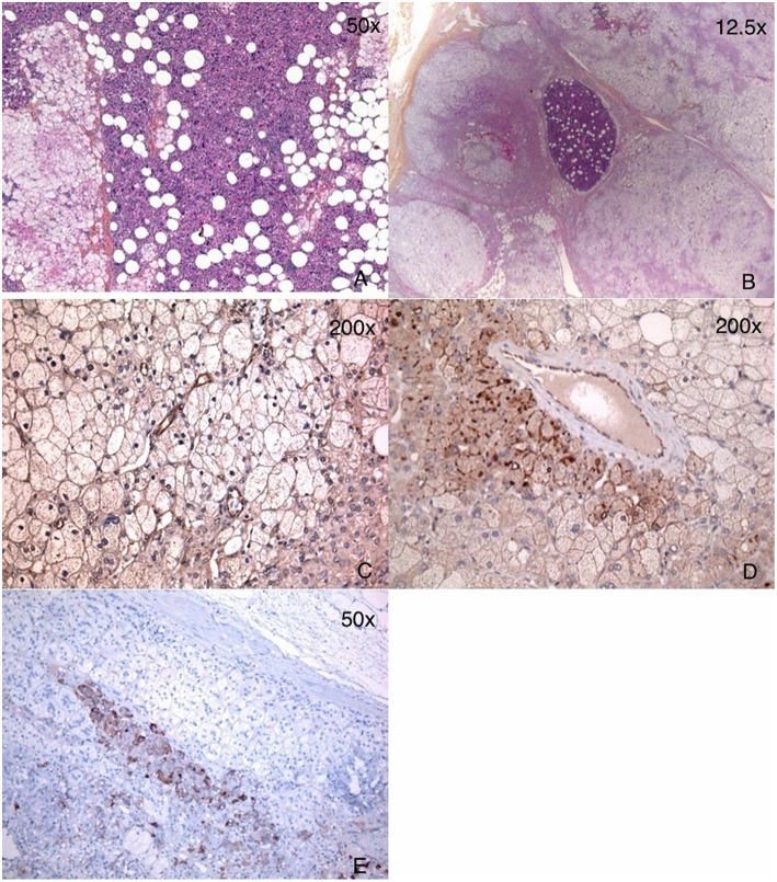
Figure 3.
Pathology of adrenal glands showing a mixture of myelolipoma and BMAH. (A) Left adrenal: large areas of myelolipoma with scattered islands of adrenocorticortical cells. (B) Right adrenal: multiple BMAH nodules and a small area of myelolipoma. (C) GIPR IHC of the left adrenal gland: only endothelial staining was seen with very minimal staining in cortical adrenal cells of BMAH. (D) GIPR IHC of the right adrenal gland: endothelial staining and focal moderate staining in membranous pattern was seen in the cortical adrenal cells of BMAH. (E) ACTH IHC showed staining only in the adrenal medulla cells, and not in BMAH cells.

