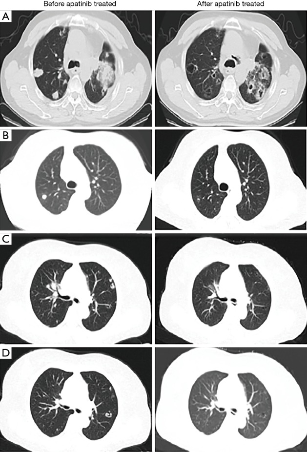
Figure 2.
Chest CT scans of patients. (A) A 60-year-old man with stage IV lung squamous cell carcinoma treated with apatinib. His baseline chest CT scan prior to therapy demonstrated a solid dominant mass in the left lung, and four nodules were noted in the right lower lobe. A follow-up CT scan at 1.5 months after apatinib therapy demonstrated that a cavity developed in the left lung within the dominant mass, decreasing in the size of the mass. The four nodules in the right lung developed cavitations. (B) A 60-year-old woman with stage IV lung adenocarcinoma who underwent right lower lobectomy 4 years ago, presenting with histologically confirmed recurrent disease in the right lung nodules. Her baseline chest CT scan prior to therapy demonstrated pleural nodularity along the right lung and small faint nodules in the left lower lobe. A follow-up CT scan at 5 months after apatinib therapy indicated the formation of a cavitation in the right lung. (C) A 52-year-old man with stage IV gastric cancer with lung metastasis who presented with histologically confirmed recurrent disease in the lung nodules. His baseline contrast chest CT scan prior to therapy demonstrated pleural nodularity along the left lung. Follow-up CT scan at 3 months after apatinib therapy indicated cavitation in these nodules. (D) A 55-year-old woman with stage IV gastric cancer with lung metastasis who presented with histologically confirmed recurrent disease in lung nodules. Her baseline contrast-enhanced chest CT scan prior to therapy demonstrated pleural nodularity along the right lung and small faint nodules in the left lower lobe. Follow-up CT scan at 7 months after apatinib therapy indicated cavitation in the nodule in the right lung.

