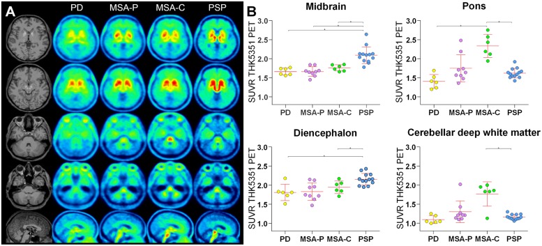
FIGURE 1.
[18F]-THK5351 tracer uptake. (A) High midbrain uptake in axial and sagittal slices allows visual discrimination of the PSP patient group from other patient groups. Significantly higher [18F]-THK5351 uptake in the diencephalon was detected in PSP patients compared to PD and MSA-C patients. In contrast, in MSA-C patients, but not in MSA-P patients, [18F]-THK5351 uptake was especially high in the pons and cerebellar deep white matter. (B) Tracer uptake in the midbrain, pons, diencephalon, and cerebellar deep white matter. ∗ indicates significant differences.

