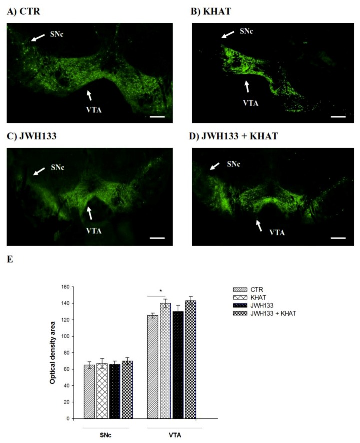
Figure 5.
Effect of sub-acute administration of khat extract alone and in combination with JWH133 on immunohistochemical staining for TH-positive cells in WT mice. (A–D) Representative photomicrographs of TH immunoreactive neurons in the substantia nigra pars compacta (SNc) and ventral tegmental area (VTA) region. Scale bar, 100 μm. (E) The number TH positive cells (optical density area), and the data were expressed as mean ± SEM (n = 6 in each group). Statistical analysis was done using one-way ANOVA followed by Tukey′s multiple comparison test. * p < 0.05. CTR (control group received the vehicle); KHAT (treatment group received khat extract 300 mg/kg); JWH133 (treatment group received JWH133 5 mg/kg); JWH133 + KHAT (treatment group received a combination of JWH133 5 mg/kg with khat extract 300 mg/kg). The vehicle, JWH133, and khat extract were administered to mice once daily for seven consecutive days.

