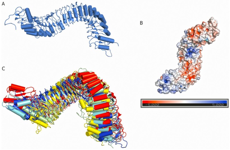
Figure 3.
Structure of an LRR domain with a twisted superhelical assembly: (A) Ribbon diagram showing the superhelical arrangement of the TDR receptor ectodomain (PDB ID 5JFK) colored in blue. (B) The TDR receptor ectodomain is shown in a surface model colored by electrostatic potential. Blue color denotes positively charged surface patches; red means negatively charged surface; and white is neutral surface regions. (C) Structural superposition of five different LRR ectodomains; FLS2 is shown in red (PDB ID 4MNA), BRI1 in pale-green (PDB ID: 3RGZ), TDR is in blue (PDB ID 5JFK), PSKR1 in yellow (PDB ID: 4Z63), and PEPR1 (PDB ID 5GR8) is denoted by cyan.

