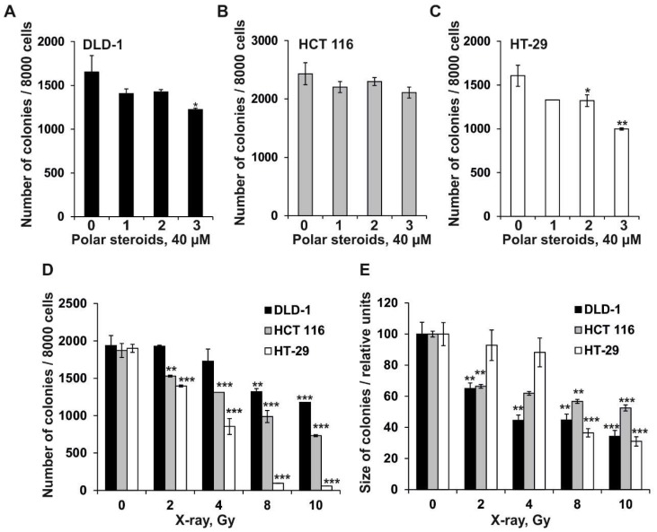
Figure 2.
The effect of polar steroids from P. pectinifera (1–3) and X-ray radiation on colony formation in human colorectal carcinoma cells. DLD-1 (A), HCT 116 (B), and HT-29 (C) cells (2.4 × 10⁴) with or without polar steroid 1–3 (40 µM) or (D,E) X-ray radiation (2–10 Gy) treatment were subcultured onto 0.3% Basal Medium Eagle (BME) agar containing 10% FBS, 2 mM L-glutamine, and 25 µg/mL gentamicin. After 14 days of incubation, the number (A–D) and size (E) of the colonies were evaluated under a microscope with the aid of the ImageJ software program. All experiments were repeated at least three times in each group (n = 9 for control or compounds treated cells or X-ray exposed cells, n—quantity of photos). Results are expressed as the mean ± standard deviation (SD). The asterisk (*) indicates a significant decrease in the number or size of the colonies of cancer cells treated by polar steroids or X-ray compared to PBS-treated cells (*p < 0.05, **p < 0.01, ***p < 0.001).

