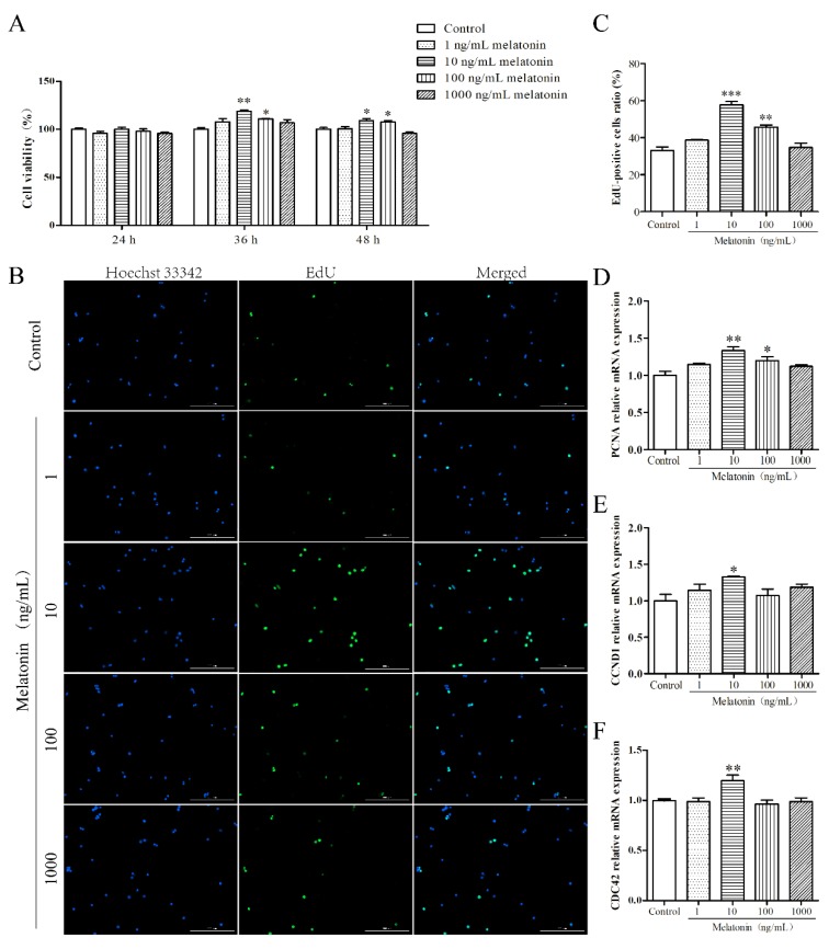
Figure 1.
Effects of melatonin on proliferation of mouse Leydig cells. (A) The effects of different concentrations (1, 10, 100, and 1000 ng/mL) of melatonin on the cell viability of mouse Leydig cells at various times (24, 48, and 72 h) (n = 3). (B) Proliferation of mouse Leydig cells treated with different concentrations of melatonin was measured using the EdU incorporation assay (n = 3). Green fluorescence represents EdU-labeled Leydig cells (original magnification ×10). (C) The proportion of EdU-positive Leydig cells as shown in panel (B). The relative mRNA expression levels of proliferating cell nuclear antigen (PCNA) (D), cyclin D1 (CCND1) (E), and cell division control protein 42 (CDC42) (F) (n = 3). Values are shown as mean ± SEM. *** p < 0.001, ** p < 0.01 or * p < 0.05 compared with the control group.

