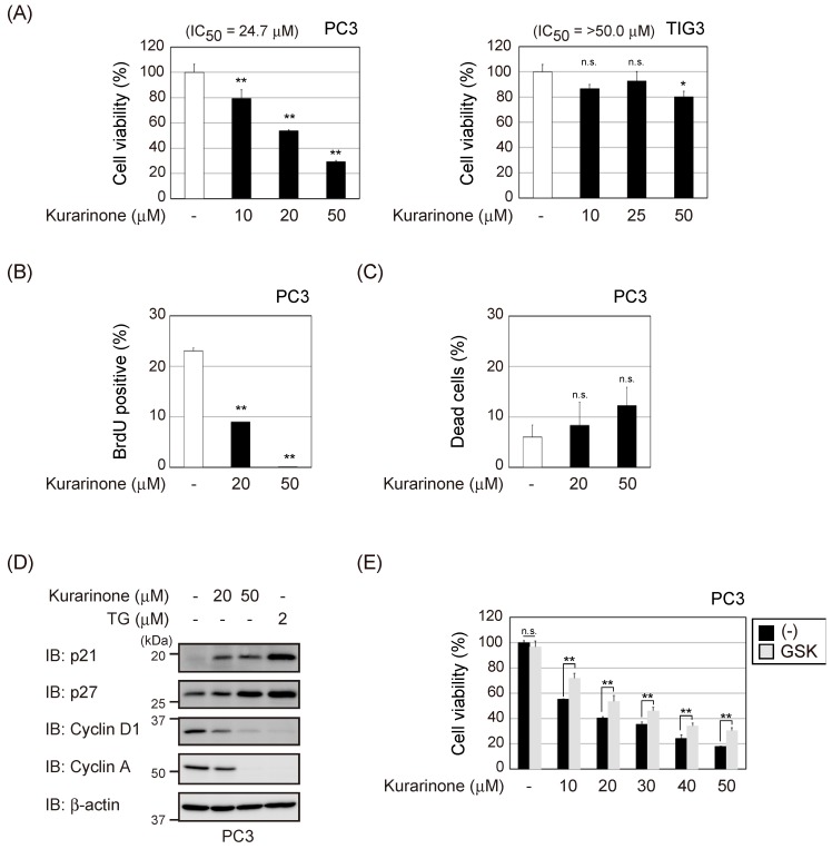
Figure 4.
Kurarinone exerts cytostatic effects on cancer cells. (A) PC3 and TIG3 cells were incubated with the indicated doses of kurarinone for 48 h. Cell viability was determined by WST-8 assay. Results represent the mean ± S.D. (n = 3). (B) PC3 cells were incubated with the indicated doses of kurarinone for 48 h. Cells were labeled with 10 μM of BrdU for 1 h and analyzed by fluorescence-activated cell sorting. The average percentage of BrdU-positive cells is shown. Results represent the mean ± S.D. (n = 3). (C) PC3 cells were exposed to the indicated doses of kurarinone for 48 h. The percentage of dead cells was measured by trypan blue staining. Results were shown as the mean ± S.D. (n = 3). (D) PC3 cells were incubated with the indicated doses of TG or kurarinone for 6 h. Cell lysates were immunoblotted with the indicated antibodies. (E) PC3 and TIG3 cells were exposed to the indicated doses of kurarinone with or without 1 μM of GSK2656157 (GSK) for 48 h. Cell viability was determined by WST-8 assay as in (A). Results represent the mean ± S.D. (n = 3). Significant differences are indicated as ** p < 0.01. * p < 0.05. n.s.: not significant.

