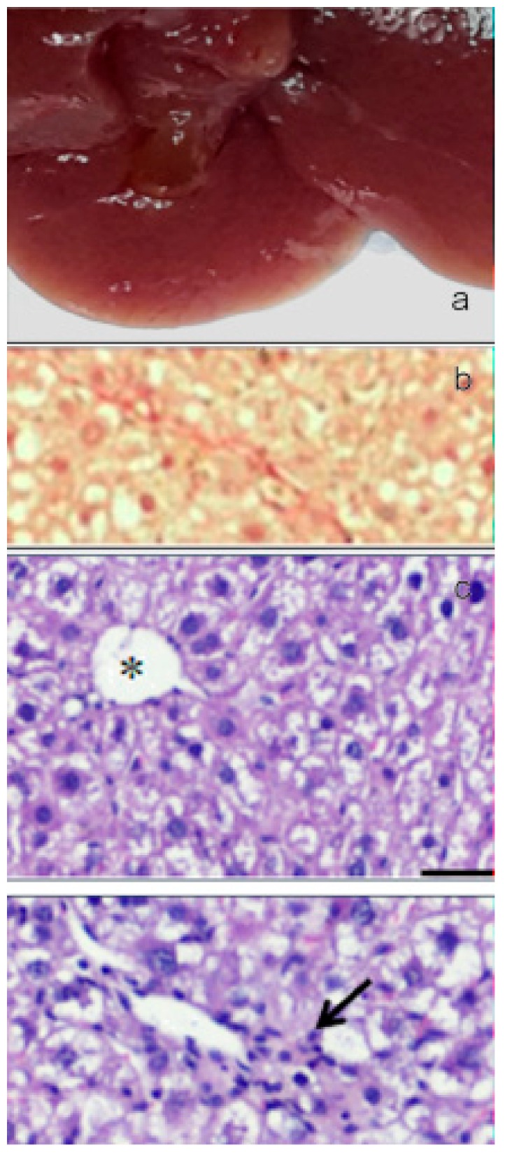
Figure 14.

(a) Image of the gross morphology of the liver showing an improvement of the pathological changes induced by CCl4 challenge in the CCl4+paramylon nanofibers group in which the normal tissue consistency was almost restored. (b) Picrosirius red staining of liver sections: reduction of the amount of collagen deposition respect to the challenged group by paramylon nanofibers treatment. (c) Reversal of severe inflammation and vein congestion along with a milder vacuolization of hepatocytes (upper image). The infiltration of inflammatory cells, although still present, is greatly reduced (bottom image, arrow). Asterisk indicates the central vein and star indicates the portal vein. Bar 50 μm.
