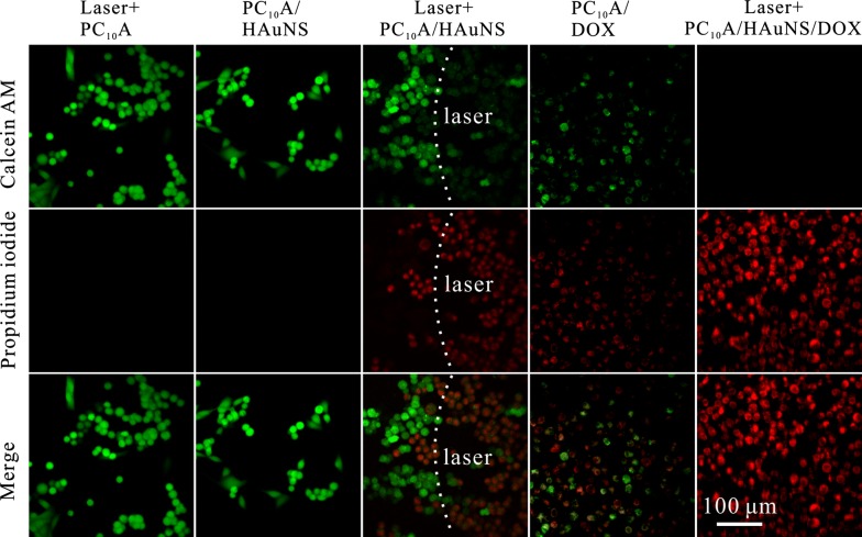
Fig. 7.
Confocal fluorescence images of HepG2 cells suffered with different treatments and stained with calcein AM/EthD-1 homodimer. The cells in PC10A hydrogel, PC10A/HAuNS hydrogel, and PC10A/DOX/HAuNS hydrogel were exposed under an 808 nm laser with a power density of 2.0 W cm−2 for 9 min (PC10A: 3% w/w; DOX: 0.8 mg mL−1, HAuNS: 20 μg mL−1)

