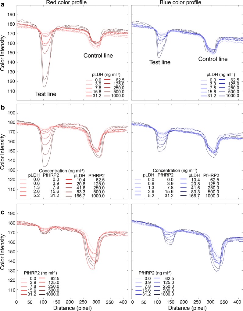
Fig. 3.
Red and blue intensity profiles of test strips. RGB colour profiles were obtained from the strip images from Fig. 2. For simplicity, red and blue intensities were represented, except for the green intensity. a When blue colours appeared on the test lines, red intensity was more decayed than blue intensity. This trend was because the background of the white strip retained high RGB values. b When mixture colour appeared at the test lines, red and blue peaks were generated corresponding to the concentration of antigens. c For PfHRP2 detection only, the blue peaks were more decayed than red peaks, in reverse observation from a

