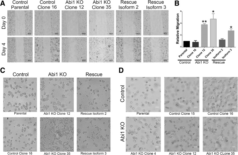
Fig. 4.
ABI1 KO cells have increased migratory ability in 2D and 3D cultures. (a) Representative images from wound healing (scratch-wound) assays at 0 days and 4 days post-scratch demonstrating increased migration of the ABI1 KO cells. Scale bars represent 200 μm. (b) The covered area in images was quantified using ImageJ. The graph represents data from n = 3 experiments (with 3 independent fields per cell line per experiment) normalized to the parental RWPE-1 cell data. The graph was generated with Prism software, and the results are represented as the mean ± SEM, unpaired Student’s t-test; * (p < 0.05); ** (p < 0.01). (c) Representative images demonstrating altered growth characteristics evidenced by the appearance of the cells in a Matrigel overlay assay. Cells were plated on a chamber slide and overlaid with Matrigel the next day. Images were taken 4 days post-overlay. ABI1 KO cells grew in a 2D culture-like manner (second column), while control RWPE-1 (first column) and ABI1 overexpression cells (third column) primarily grew into spheroids. (d) When plated in regular 3D cultures, the ABI1 KO organoids showed invasive/migratory abilities. The images show parental cells, two failed CRISPR control clones (top row) and three different ABI1 KO clones (bottom row) at 4 days post-culture. Scale bars represent 100 μm

