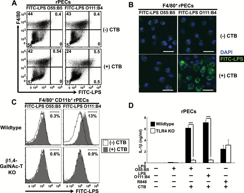
Fig. 2.
CTB induces IL-1β production from rPECs prestimulated with LPS O55:B5, which fails to enter the cytosol of peritoneal macrophages, in a TLR4-dependent manner. (A–C) Assessment of LPS internalization in rPECs. rPECs from wild-type (A) and wild-type and β1,4-GalNAc-T-deficient (C) mice were first cultured with 5 μg ml−1 FITC-conjugated LPS O55:B5 or FITC-conjugated LPS O111:B4 for 5 h and further cultured for 19 h with or without 20 μg ml−1 CTB. Cells were then harvested, stained and subjected to flow cytometry analysis. In (A), dot plots of F4/80 versus FITC-LPS from wild-type rPECs are shown. Numbers indicate percentages of gated cells among total cells. Data are representative of three independent experiments. In (C), histograms of FITC-LPS from wild-type and β1,4-GalNAc-T-deficient F4/80+ CD11b+ cells are shown. Shaded and open histograms indicate data from cells cultured with and without CTB, respectively. Numbers indicate the percentages of gated cells among total cells. Data are representative of three independent experiments. In (B), F4/80+ rPECs cultured with or without CTB were subjected to confocal microscopic analysis. Green and blue colors represent LPS internalization and DAPI staining, respectively. The scale bars represent 20 μm. Data are representative of two independent experiments. (D) rPECs from wild-type C57BL/6 or TLR4-deficient mice were first cultured for 5 h in the absence or presence of 500 ng ml−1 LPS O55:B5, 500 ng ml−1 LPS O111:B4 or 100 nM R848. Then 20 μg ml−1 CTB was added and further cultured for 19 h. IL-1β production was measured by ELISA. Data are representative of two independent experiments. The results are presented as means ± SD. ***P < 0.001.

