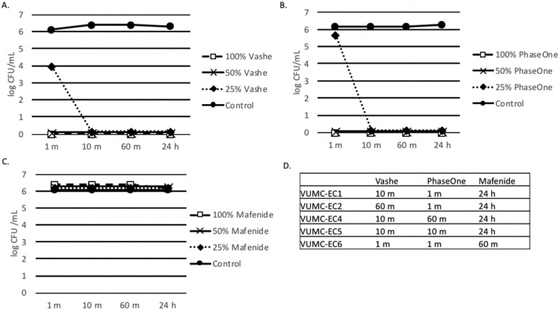
Figure 3. Effect of Wound Solutions on E. coli.
A-C. Biofilms were formed in 100 μl BHI with 1% glucose. E. coli ATCC 25922 (panels A-C) or clinical strains of E. coli (panel D). Organisms were added to tissue culture treated plates and incubated for 24 h at 35°C. After 24 h biofilms were washed with 1X PBS three times and 100 μl 100%, 50% or 25% Vashe (panel A), PhaseOne (panel B) or Mafenide (panel C) were added to the wells. Control wells contained 100 μl 1X PBS. Plates were incubated for 1 m, 10 m, 60 m or 24 h. Wells were washed three times with PBS to remove the wound solutions and reconstituted with 100 μl H2O. Bacterial viability was monitored by the CFU assay. D. This data represents the minimum time to kill (CFU=0) clinical strains of E. coli with Vashe, PhaseOne or Mafenide. Bacteria that were not killed at 24 h are indicated by >24 h.
Panels A-D are representative of 3 independent experiments.

