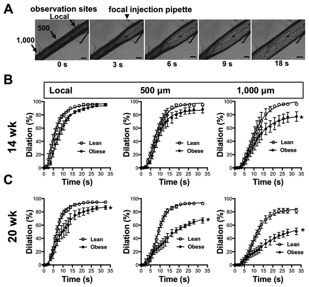
Figure 2. Impaired conducted vasodilation in obese ZSF1 rats.
A; Representative time lapse images show conducted vasodilation in an isolated pressurized skeletal muscle artery of the obese ZSF1 rat. Panels B and C: Summary data of conducted vasodilation in skeletal muscle artery in response to focally applied acetylcholine at local and upstream sites (500 and 1,000 μm) over time in lean and obese ZSF1 rats at age of 14 weeks (B; n=4-4) and 20 weeks (C; n= 5-6). * indicates P < 0.05 lean versus obese ZSF1 rats.

