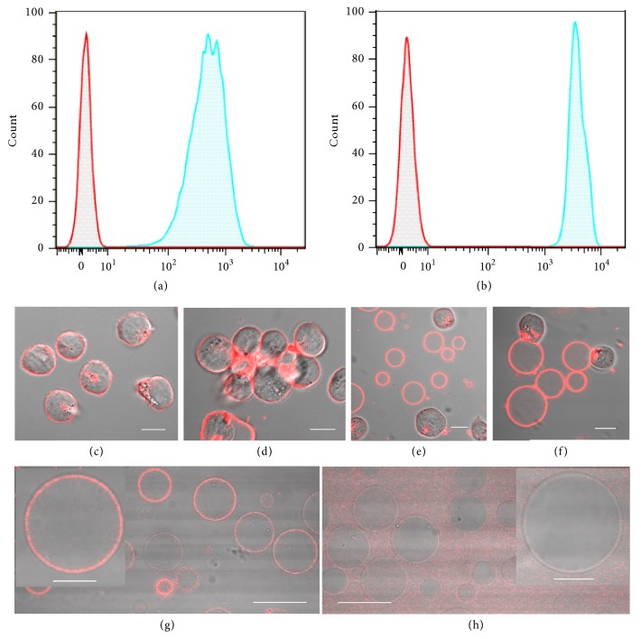
Figure 2.
GMVs exhibit well-preserved membrane properties. Flow cytometry analysis of (a) GMVs and (b) HeLa cells incubated with fluorescein-labeled WGA. Confocal images of CCRF-CEM cells and GMVs demonstrate the similar response to WGA-induced aggregation. Overlay images of bright field and fluorescent images of CCRF-CEM cells (c) before and (d) after incubation with WGA. Mixture of CCRF-CEM cells and GMVs (e) before and (f) after incubation with WGA. The overlay images of GMVs incubated with PE-labeled Mouse anti-Human CD71 antibody (g) or Mouse IgG 2a (h) demonstrate the good recognition capability of CD71 on GMVs. About 10000 events were counted for each sample. Scale bar is 30 μm for (g), (h) and 10 μm for (c)-(f) and the inset figures in (g) and (h).

