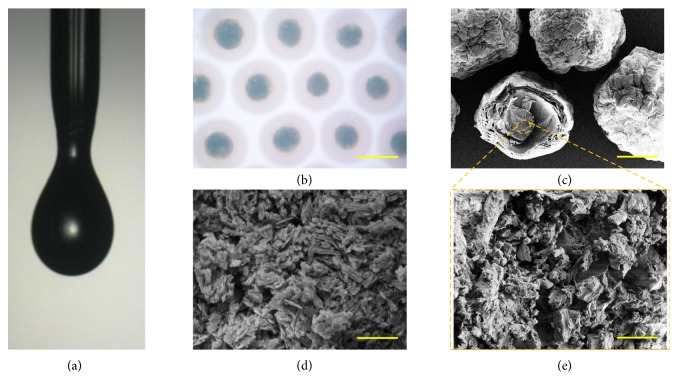
Figure 2.
The preparation of the niacin Cu-MOFs encapsulated microcapsules. (a) The real-time image of the microfluidic electrospray process of the microcapsules. (b) Microscope image of the microcapsules. (c–e) Scanning electron microscope (SEM) images of the (c) niacin Cu-MOFs encapsulated microcapsules, (d) niacin Cu-MOFs, and (e) niacin Cu-MOFs inside the microcapsules. Scale bar in (b) is 300μm, in (c) is 100μm, and in (d, e) is 5μm.

