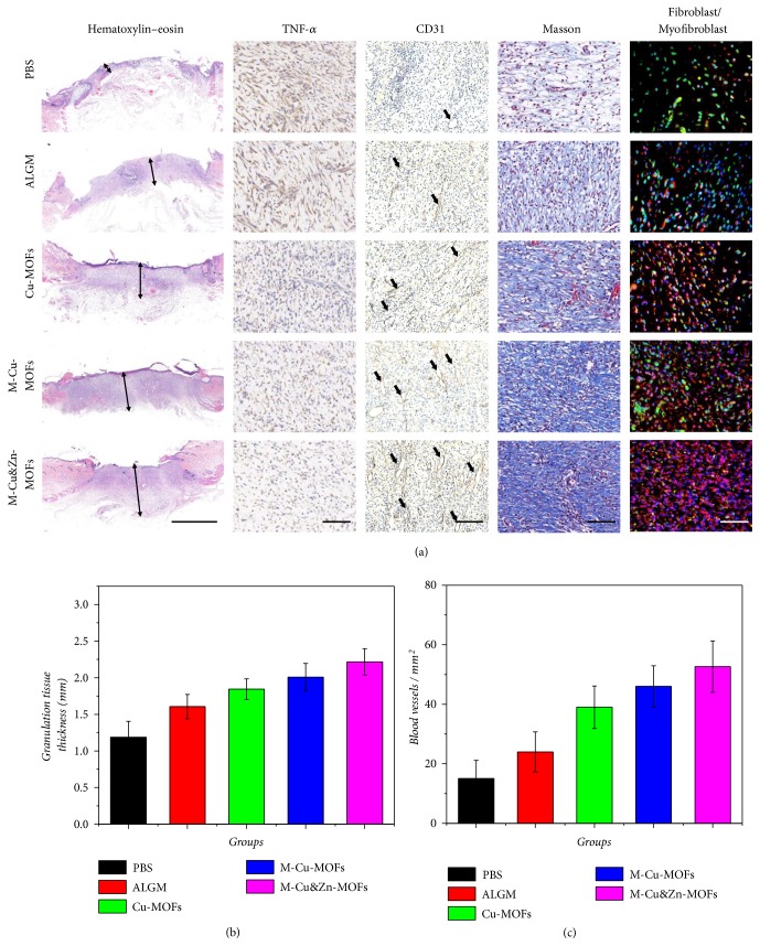
Figure 6.
Biological mechanism of the wound healing process. (a) Hematoxylin-eosin staining, IHC staining of TNF-α and CD31, Masson trichrome stain, and double immunofluorescent staining of fibroblast marker vimentin (green) and myofibroblast marker α-SMA (red) of granulation tissues in the wound bed after 7 d at low magnification. (b) Quantitative analysis of granulation tissue thickness. (c) Quantification of CD31 labeled structures. Scale bar in (a) is 2mm in hematoxylin-eosin staining, 100μm in IHC staining of TNF-α, Masson trichrome stain, and double immunofluorescent staining, and 200μm in IHC staining of CD31.

