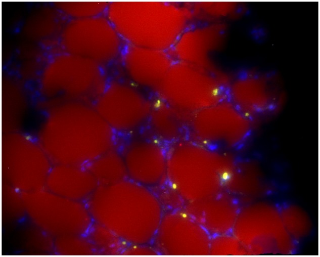
Figure 1.

GFP+ 8093 acute lymphoblastic leukemia cells in the perirenal fat pad of a transplanted obese mouse that developed progressive leukemia during vincristine treatment. Lipid is stained with Nile Red, and the image is counterstained with 4', 6-diamidino-2-phenylindole. Image was taken on a Leica DM RXA2 with a x20 objective with x1.6 Optovar magnification (x32 final). Image is representative of fat pads obtained from the other four mice after vincristine treatment. Reprinted with permission from (5).
