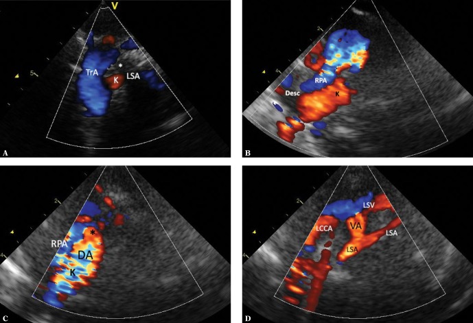
Fig. 10.
RAA ALSA Kommerell’s diverticulum. A. A horizontal mediastinal section showing the right aortic arch (TrA) and its final branch – the left subclavian artery (LSA) located retrotracheally in its initial segment (*), which makes it very difficult to visualize its course. B. A slight parallel downward movement of the transducer allows for more precise visualization of the initial, wide segment of the left subclavian artery (Kommerell’s diverticulum – K) arising from the distal portion of the aortic arch (Desc; it runs approximately parallel, slightly above the right pulmonary artery – RPA). Furthermore, a transverse section of the pulmonary trunk is visible. C. The ductus arteriosus (DA) is still patent, representing a nearly smooth continuation of the Kommerell’s diverticulum (K). The ductal orifice to the pulmonary trunk (*) and a fragment of the right pulmonary artery (RPA) are visible. D. A parallel leftward movement of the transducer shows the further course of the LSA, which becomes significantly narrower after forming the ductus. A division into the distal LSA and the vertebral artery (VA) is seen. The left common carotid artery (LCCA) and the left subclavian vein (LSA) are visible anteriorly to the LSA

