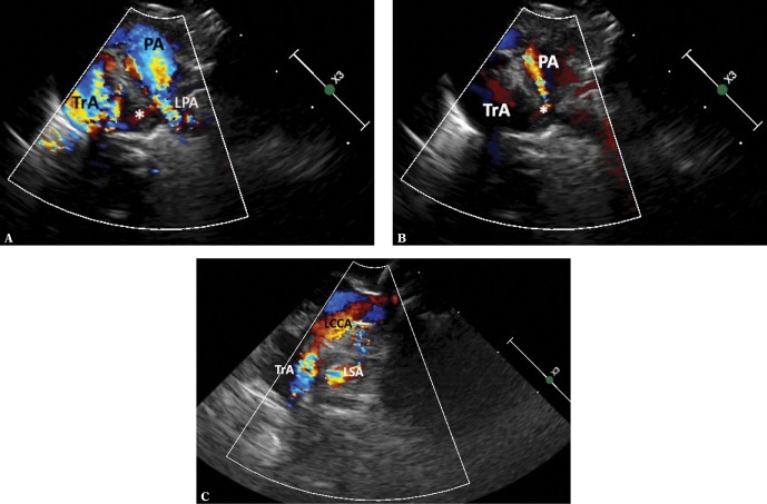
Fig. 11.
RAA with an aberrant subclavian artery, the ductus arteriosus arising directly from the descending aorta and connecting to the LPA. A. A high parasternal view showing the upper mediastinum in a transverse plane. Systole. Visible elements: the pulmonary artery trunk (PA), its left branch (LPA), distal transverse portion of the aortic arch (TrA). These vessels are coded blue. Red-coded flow in the ductus arteriosus (*) is seen between the descending aorta and the initial segment of the left pulmonary artery. The vascular structures form a complete vascular ring around the trachea and esophagus. B. View and legends as in Fig. 11 A. Diastole. Ductus arteriosus shunt into the initial segment of the left pulmonary artery and the pulmonary trunk is more visible during this phase of the cardiac cycle. C. A slight upward movement of the ultrasound beam visualizes both left-sided branches of the aortic arch: the left common carotid artery (LCCA) and the left subclavian artery (LSA), which arises separately from the distal part of the aortic arch

