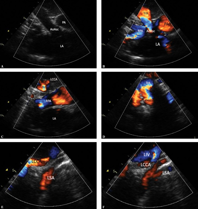
Fig. 6.
Imaging of the right aortic arch. A. A suprasternal view, mediastinal section in the frontal plane. Diastolic phase. Elements visualized: ascending aorta (AoAsc), pulmonary artery trunk (PA), left atrium (LA). B. The view and legends as in Fig. 5A, a slight posterior tilt of the ultrasound beam, systole. Elements that are still visible: ascending aorta up to the apex of the transverse aortic arch, pulmonary trunk, the right pulmonary artery crossing the ascending aorta, with its division into upper lobar artery, middle artery, and the left atrium. C. Further posterior movement of the ultrasound beam no longer shows the aorta; the right pulmonary artery and the pulmonary trunk, as well as the transverse part of the aortic arch along with the first, leftward branch are still visible. It is difficult to decide in this view whether this is the left brachiocephalic trunk or the left subclavian artery. D. A definite backward tilting of the transducer shows the right aortic arch with its convexity directed to the right and the initial segment of the descending aorta. E. A wide, strong tracheal shadow significantly obscures the descending aorta; therefore, precise assessment of its lower segment in the image from the previous figure is not possible. Slight parallel movement of the transducer to the left visualizes both the longer segment of the first arch branch – a relatively narrow and arched upwards left common carotid artery (LCCA) – as well as the left subclavian artery (LSA) emerging from the tracheal acoustic shadow, initially wide and narrowing towards the periphery. The image is indicative of the presence of an aberrant left subclavian artery, arising from the descending aorta. F. Continued movement to the left allows following further course of the left branches of the aortic arch. LIV – left innominate vein

