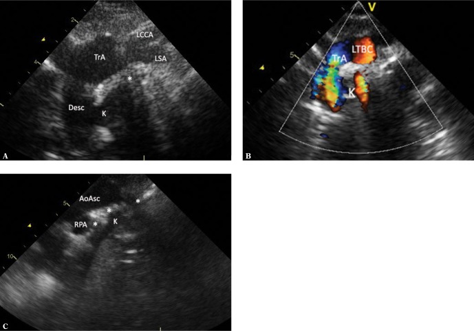
Fig. 8.
RAA with brachiocephalic trunk, the ductus arteriosus arising from the descending aorta and passing to LPA. A. A view showing the transverse portion of the aortic arch (TrA) with a clear rightward convexity. A very wide and short left brachiocephalic trunk dividing into a horizontal left subclavian artery (LSA) and the left common carotid artery (LCCA), which bends upwards. The distal part of the aortic arch is deformed by a wide Kommerell’s diverticulum (K), which is largely obscured by the trachea (*). B. A view showing the right aortic arch in a similar way as in Fig. 1A. Color-coded flow; however, branches of the aortic arch are not visible. C. A view showing the ascending aorta (AoAsc) in a sagittal plane. The trachea (*) is located just behind the aorta, slightly above the crossing with the right pulmonary artery (RPA), significantly deformed by a hyperechoic structure – the Kommerell’s diverticulum (K)

