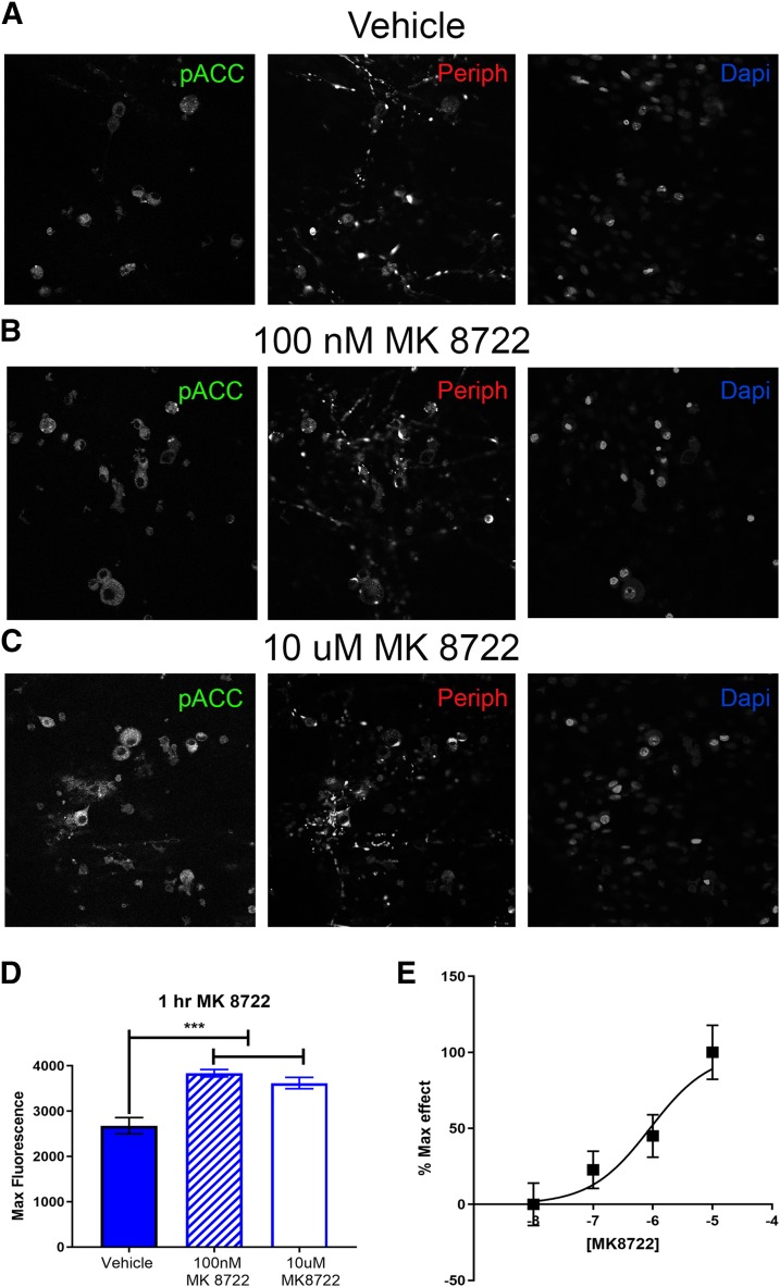
Fig. 9.
MK8722 induces AMPK activation in DRG neurons in vitro. Neuron cultures were treated with vehicle (A), 100 nM (B), or 10 µM MK 8722 (C) for 1 hour Representative immunohistochemistry images of the DRG neurons at 40× magnification. Quantification of images shown in (D). 100 nM and 10 µM NCLS increased p-ACC intensity in male neuron cultures. Only neurons positive for peripherin staining were analyzed. Maximum florescence refers to the maximum florescence intensity per neuron analyzed. **P < 0.01; ***P < 0.001. n = 39 images analyzed per group. (E) Shows a full concentration-response curve of MK8722 with an approximate EC50 of 900 nM (95% confidence interval, 253 nM–2.62 μM).

