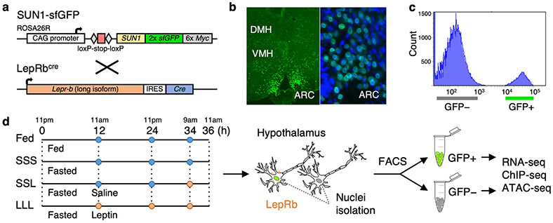
Fig. 1.
Isolation of LepRb GFP-positive nuclei. a R26-CAG-LSL-Sun1-sfGFP-myc homozygous mice were crossed with LepRbcre mice to obtain mice that express sfGFP in LepRb expressing cell nuclei. b Immunostaining showing sfGFP expression in the dorsomedial hypothalamus (DMH), ventromedial hypothalamus (VMH) and arcuate nucleus (ARC). On the right panel is a zoom in on the ARC along with DAPI staining (blue). c FACS histogram showing clear separation of GFP-positive from GFP-negative nuclei obtained from LepRbcre X R26-CAG-LSL-Sun1-sfGFP-myc heterozygous mice. Similar results were obtained in all the experimental groups (Supplementary Figure 1). d LepRbcre X R26-CAG-LSL-Sun1-sfGFP-myc heterozygous mice were fed or fasted for 36 hours, during which saline (blue) or leptin (orange) were injected at various time points. Cell nuclei were isolated and sorted via FACS, followed by RNA-seq, ChIP-seq, and ATAC-seq.

