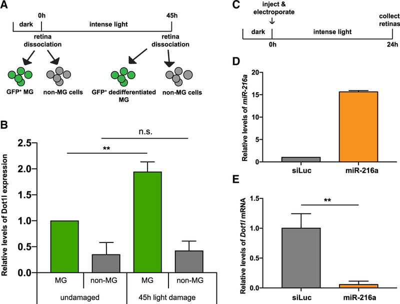
Figure 3. miR-216 Targets Dot1l in the Retina during Photoreceptor Regeneration.
(A) For post-mitotic MG isolation, GFP+ cells were sorted from dark adapted undamaged Tg(gfap:gfp) retinas. For dedifferentiated MG isolation, GFP+ cells were isolated from 45-h light-lesioned Tg(1016tuba1a:GFP) retinas.
(B) Fold changes in dot1l levels in FACS-purified MG were determined by qPCR. After 45 h of light damage, dot1l is upregulated in dedifferentiated MG (GFP+) in Tg(1016tuba1a:gfp) fish. Dot1l expression did not change in non-MG cells (GFP−) during regeneration. Data represent the means ± SEMs from 15 undamaged fish, and dedifferentiated MG were purified from 18 light-damaged fish.
(C) Experimental scheme to test the effects of miR-216a overexpression on dot1l levels. Wild-type adult zebrafish were dark adapted and then either control miRNA or miR-216a was injected and electroporated into the left eyes before intense light exposure. After 24 h of light exposure, retinas were dissected for RNA isolation.
(D) Fold changes in miR-216a levels in control miRNA (small interfering RNA [siRNA] against luciferase [siLuc]) or miR-216a mimic electroporated retinas were quantified by qPCR. miR-216a levels were upregulated by 15-fold in miR-216a mimic-injected retinas compared to controls. Data represent the means ± SEMs from 6 retinas.
(E) Fold changes in dot1l levels in siLuc or miR-216 mimic electroporated retinas were quantified by qPCR. After 24 h of light damage, dot1l was downregulated in miR-216a-overexpressing retinas ~20-fold. Data represent means ± SEMs from 3 independent experiments. Six retinas were pooled for RNA isolation in each experiment.
**p < 0.01 (Student’s t test), p = 0.0093.
Scale bars, 50 µm.
See also Figures S1 and S2.

