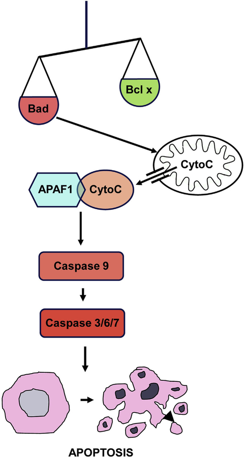
Fig. 2. Diagram of the intrinsic pathway of apoptosis.
Apoptosis can be initiated when pro-apoptotic proteins such as BAX or BAD which overwhelm anti-apoptotic proteins of the BCL family. This destabilizes mitochondrial membranes, releasing Cytochrome c into the cytoplasm. Interaction of Cytochrome c with APAF forms an apoptosome, which enzymatically cleaves pro-caspase 1 into its active form. This initiator caspase in turn cleaves executor caspases (3, 6 and 7), which mediate programmed death and orderly fragmentation of the cell.

