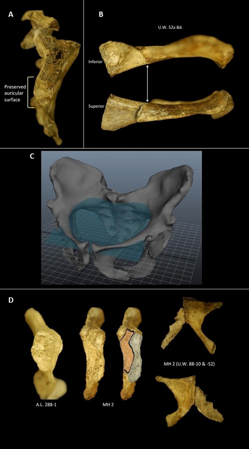
Fig 2. Detailed anatomy of the MH2 pelvis that informed the reconstruction used in this study.

A. Preserved auricular surface on the MH2 sacrum. B. Superior pubis showing tight, direct contact between U.W. 88–52 and U.W. 88–136 (previously called U.W. 88-52b). C. Plane fitted to composite pelvis showing the arcuate line is continuous. D. Pubis of MH2 (U.W. 88–52 and U.W. 88–10) articulated. E. Comparison of the pubic symphysis between A. afarensis (A.L. 288–1) and A. sediba (MH2). Outlined in gray is the articular surface for the contralateral pubis; outlined in orange is the non-articular portion of the symphysis and presumed insertion for the anterior pubic ligament.
