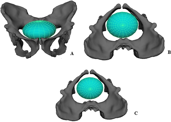
Fig 4. Ellipse representing a neonatal A. sediba head at the pelvic.
A. inlet, frontal view B. inlet, superior view C. midplane, superior view. Reconstructed pelvis is shown with the MH1 ischium. Notice that the modeled A. sediba neonatal cranium can descend into the midplane without bony constraints, unlike the condition typically found in modern humans.

