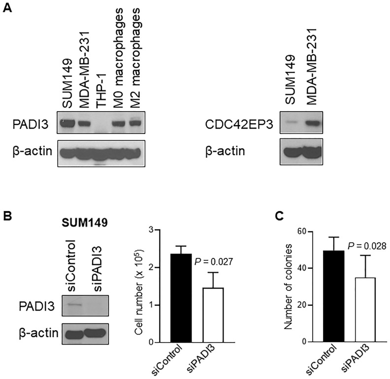
Fig 3. Analysis of PADI3 and CDC42EP3.
(A) The expression of PADI3 and CDC42EP3 in breast cancer cell lines and macrophages. Expressions of PADI3 in SUM149 (TN-IBC), MDA-MB-231 (TN-non-IBC) cells, THP-1 (monocytes), M0 macrophages (THP-1-derived immature macrophages) and M2 macrophages (THP-1–derived M2 macrophages) were analyzed with Western blotting. Expressions of CDC42EP3 in SUM149 and MDA-MB231 were analyzed with Western blotting. (B) PADI3 knockdown suppresses cell growth in SUM149. Left panel: The expression of PADI3 in SUM149 cells was depleted with siRNA and the expression of PADI3 in siControl and siPADI3 was analyzed with western blot. Right panel: Proliferation of SUM149 cells transfected with siControl and siPADI3 was measured by Trypan blue exclusion assay. Bars, SD. (C) PADI3 knockdown suppresses anchorage-independent growth of SUM149 cells. Bars, SD.

