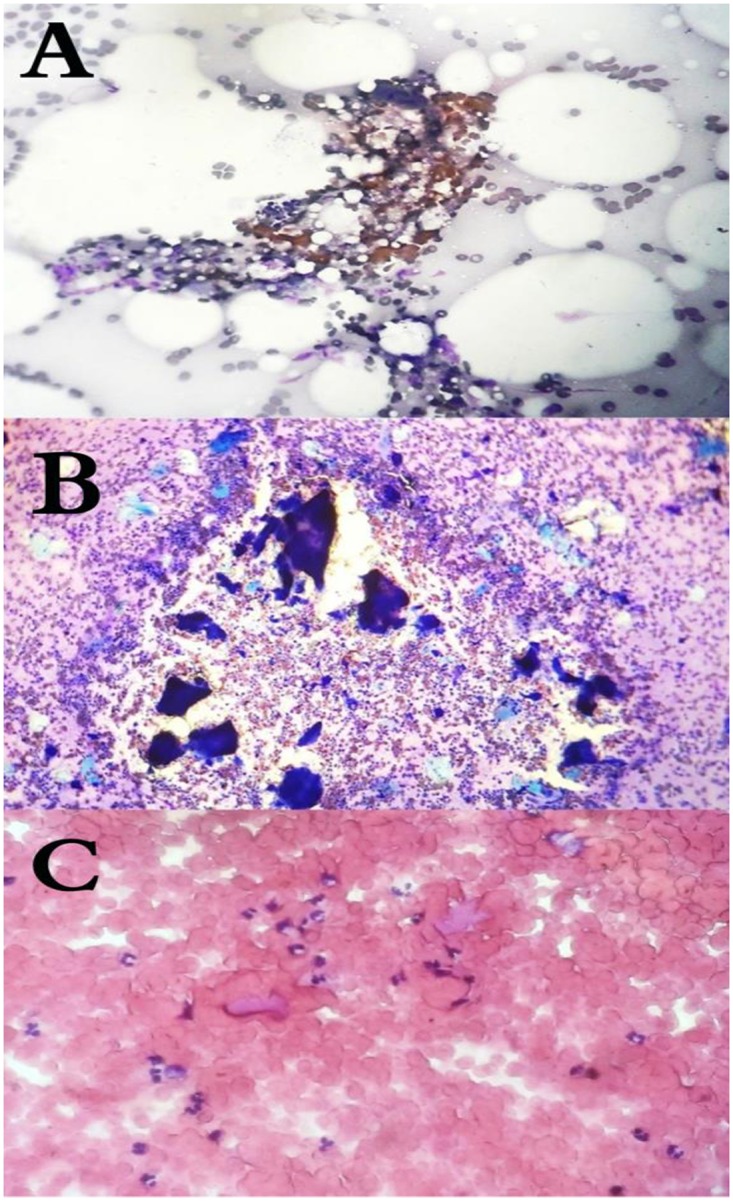
Fig 2. Cytology of mycetoma causative agents.

(A): The cytological appearance of M. mycetomatis, (B): A. pelletierii, (C): S. somaliensis in fine needle aspirates stained with HE (magnification 10 times).

(A): The cytological appearance of M. mycetomatis, (B): A. pelletierii, (C): S. somaliensis in fine needle aspirates stained with HE (magnification 10 times).