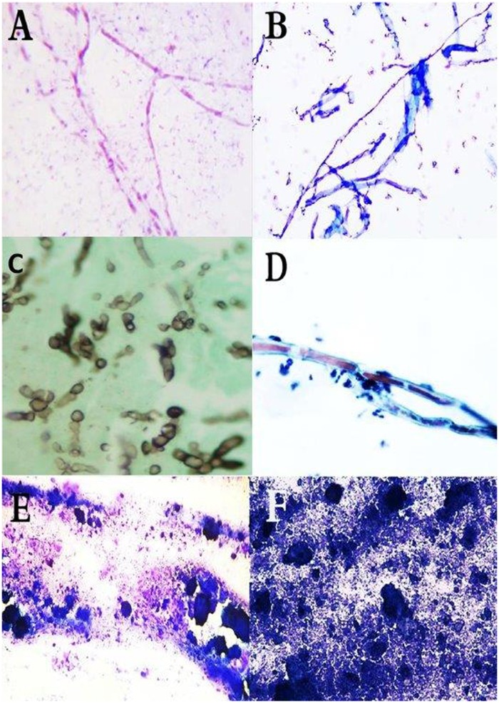
Fig 5. False positive identifications in cytology.
Showing cytological smears of (A) M. mycetomatis hyphae stained with Giemsa stain, (B) Synthetic Fibers stained Wright-Giemsa stain. (Giemsa stain, X40). (C) Smear showing hyphae of M. mycetomatis. (D) Smear showing elongated structures of the Oedogoniales order. The chloroplasts form a chain interrupted by clear zones (X40). (E) Smear showing M. mycetomatis grains after being crushed on the smear. (F) smear with abundant calcific debris without intact cells taken from patient with tumoral calcinosis (Diff Quick, X10).

