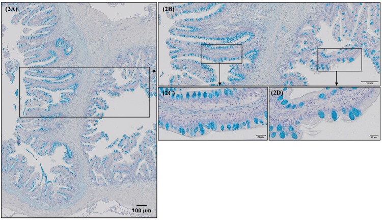
Fig 2. Morphological differences between European sea bass (Dicentrarchus labrax) posterior gut (preileorectal valve segment) and rectum (postileorectal valve segments) goblet cell size and distribution on mucosal surface.
A similar morphology and mucus production pattern were observed in all the fish intestinal sections studied. A general overview of goblet cell distribution in posterior gut and rectum (separated by ileorectal valve) is detailed in Fig 2A and 2B (Alcian Blue, pH = 2.5; Bar 100 μm). Note the greater fold area covered by mucus in posterior gut compared to rectum as a result of increased cell density as posterior gut goblet cells are smaller than goblet cells located in rectum (Fig 2C vs 2D; Alcian Blue, pH = 2.5; Bar 20 μm).

