Figure 2.
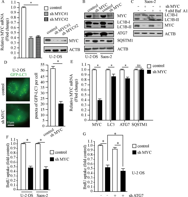
MYC promotes the proliferation of osteosarcoma cells. Cells treated with lentivirus carrying MYC shRNA or control shRNA. (A) The expression of MYC was examined by qPCR and Western blot. ACTB was used as a loading control. (B and C) Whole cell lysates were prepared, and subjected to Western blot analysis using indicated antibodies. Treatment of 5 nM Baf A1 for 4 hrs. (D) Representative fluorescence microscope images of cells stably expressing GFP-LC3, scale bar: 10 μm (left panel). Puncta of GFP-LC3 per cell (right panel). (E) Transcription levels of autophagy factors. ACTB was used as a loading control. (F and G) Cellular proliferation was assessed using BrdU assay, n=5. The data are represented as the mean ± SEM of five experiments. *P<0.01.
Abbreviations: PLK1, polo-like kinase 1; MYC, MYC proto-oncogene.

