Figure 4.
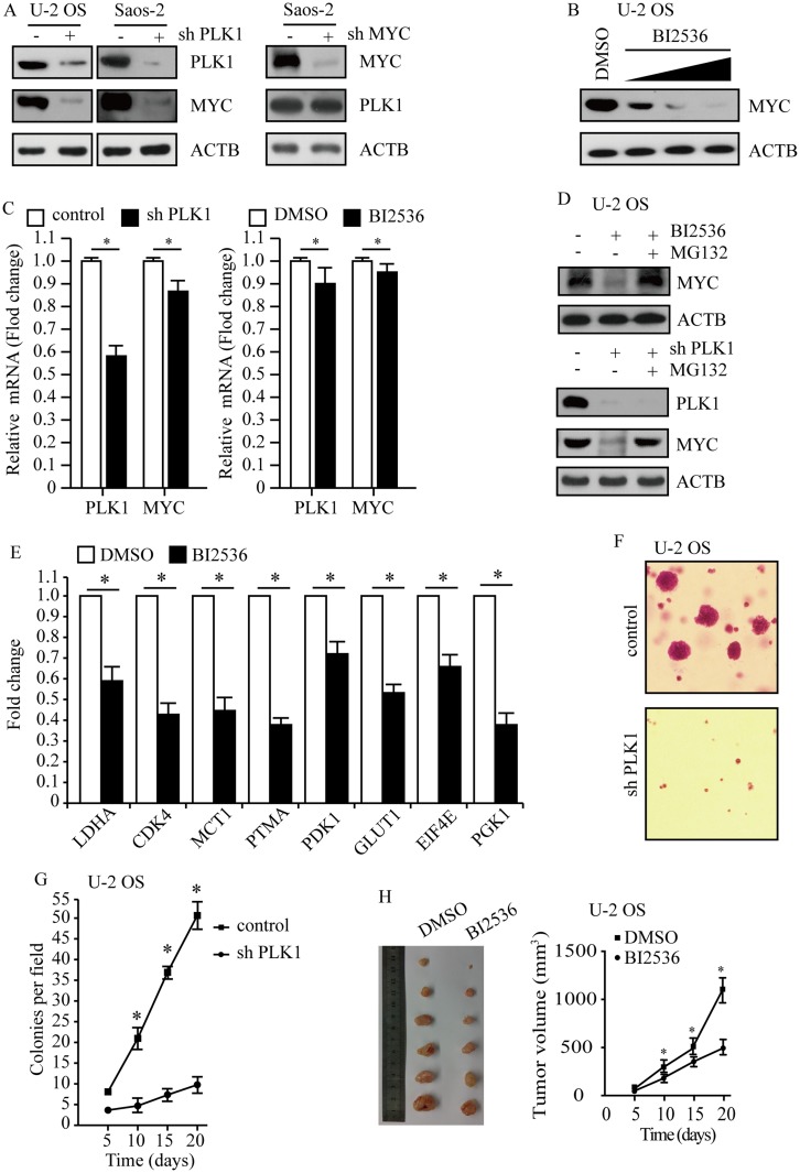
PLK1 promotes osteosarcoma development by regulating MYC stabilization. (A) PLK1 and MYC protein levels were analyzed by immunoblot, with ACTB as a loading control. (B) Immunoblot detection of MYC in U-2 OS cells after 24 hrs of treatment with BI2536 (5 nM, 15 nM 30 nM). ACTB was used as a loading control. (C) Transcription levels of PLK1 and MYC. ACTB was used as a loading control. (D) Immunoblot detection of MYC in U-2 OS cells under treatment with BI2536 (30 nM) for 24 hrs, followed by MG132 (5 μM) for another 6 hrs. ACTB was used as a loading control. (E) qPCR analysis of representative MYC target genes in U-2 OS cells upon BI2536 (30 nM) treatment for 24 hrs. Data shown represent the means (± SEM) of triplicates. (F) Clonogenic assays performed with control and PLK1 shRNA U-2 OS cells. (G) The graph shows the quantification of the mean number of colonies at different time point as indicated. (H) U-2 OS cells were subcutaneously implanted into female athymic nude mice (n=6 for each experimental condition). The tumor images on day 20 (left panel). Tumor growth curve (mean ± SEM) is shown (right panel). *P<0.01 compared to control.
Abbreviations: PLK1, polo-like kinase 1; MYC, MYC proto-oncogene.

