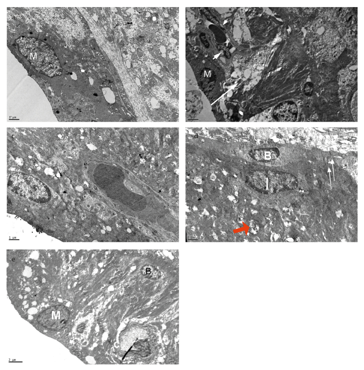
Figure 5. a–e.
Marginal cells with vacuoles and sparse basal infoldings in Group I (CDDP+IP FA). Empty spaces in the cytoplasm of intermediate cells (arrow), vacuoles in the cytoplasm of endothelial cell (arrowhead) in Group II (CDDP). Short microvilli and numerous mitochondria, basal infoldings of marginal cells and cristalysis in the mitochondria (red arrow) of Group III (CDDP+IT FA). Vacuoles in the lateral and basal foldings of marginal cells in Group V. M, marginal cell; I, intermediate cell; B, basal cell; C, capillary. Group I (CCDP+IP FA) (a), Group II (CDDP) (b), Group III (CCDP+IT FA) (c, d), Group V (IT FA) (e). X12000 (a), X6000 (b), X12000 (c), X12000 (d), and X8000 (e). Uranyl acetate-lead citrate.

