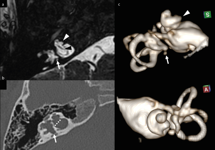
Figure 2. a–c.
Magnified axial MRI FFE T2-weighted image (a) and axial CT image (b) of right temporal bone. Volume rendering of the right internal acoustic canal and inner ear (MRI cisternography), superior and anterior view (c). Bulbous dilatation of the bottom of IAC and lack of the cribriform plate (dotted line), with direct communication between IAC and the basal turn of cochlea. The cochlea is dysmorphic (white arrowhead), with the absence of the modiolus. Note the normal cochlear nerve within IAC (black arrowhead). Also note the dysmorphic profile of the vestibule and semicircular canals, with small saccular dilatations of the wall (arrow).

