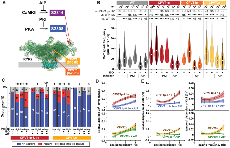
Figure 5. CaMKII inhibition suppresses the CPVT arrhythmic phenotype.

A. Schematic of RYR2 (two subunits of tetramer shown). Three key residues are highlighted: S2808, the target of PKA phosphorylation; S2814, the target of CaMKII phosphorylation; and R4651, mutated in CPVTp. B. iPSC-CMs in isolated cell clusters were treated with ISO and selective CaMKII (AIP) or PKA (PKI) inhibitors. Ca2+ sparks were imaged by confocal line scanning within individual iPSC-CMs. Data without inhibitors are the same as in Fig. 1c. Steel-Dwass non-parametric test with multiple testing correction: *, P<0.05; **, P<0.01; ***, P<0.001. NS, not significant. C. Occurrence of re-entry in CPVT1 and CPVT2 engineered tissues treated with a CaMKII inhibitor (AIP). Pearson’s chi-squared test: † vs WT with same pacing and ISO conditions; § vs. CPVT with same pacing and ISO conditions. The Bonferroni correction for multiple comparisons was applied for three possible pairwise tests performed with a corrected p threshold: †, §, P<0.05/3. ††, P<0.01/3. †††, §§§, P<0.001/3. Bars are labeled with samples sizes. D–E. Relative diastolic Ca2+ level change from basal condition (D), and spatial and temporal dispersion of Ca2+ wave propagation speed (E) as a function of pacing frequency under ISO stimulation in CPVT1 and CPVT2 before and after AIP loading. Only tissues responding 1:1 to every stimulus were included in d and e (n=33 CPVT1p, 15 CPVT1p + AIP, 13 CPVT1e, 15 CPVT2e, and 9 CPVT2e + AIP from > 3 harvests).
