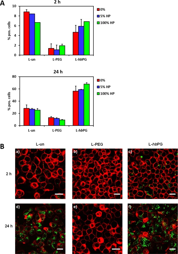
Figure 6.
(A) Influence of protein corona formation on the cellular uptake behavior of liposomes. Liposomes were either directly incubated with RAW 264.7 cells (referred to as 0%) or preincubated with human plasma (5 or 100%) and further added to cells at a concentration of 7.5 μg mL–1. Cellular interaction was analyzed by flow cytometry after 2 and 24 h. The amount of fluorescence-positive cells (%) is shown. (B) Representative CLSM images. Liposomes were treated with 100% human plasma and incubated with RAW264.7 cells for 2 or 24 h at a concentration of 75 μg mL–1. The cell membrane was stained with CellMask Deep Red and is pseudocolored in red. Liposomes are pseudocolored in green. Scale bar: 20 μm.

