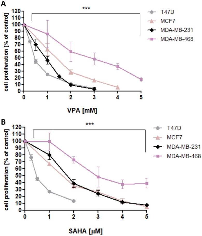
Figure 1.
Effect of VPA (A) and SAHA (B) on proliferation of T47D, MCF7, MDA-MB-231 and MDA-MB-468 breast cancer cells in Brdassay.
Notes: T47D, MCF7, MDA-MB-231 and MDA-MB-468 cells were incubated for 48 hrs alone (control) or in the presence of VPA (0.25-10 mM) or SAHA (0.25-5 µM). The differences between groups were evaluated using the one-way ANOVA; Tukey’s post-hoc test. p<0.05 was considered to indicate a statistically significant difference. Results were presented as mean ± SD of the mean.
Abbreviations: SAHA, vorinostat; VPA, valproic acid.

