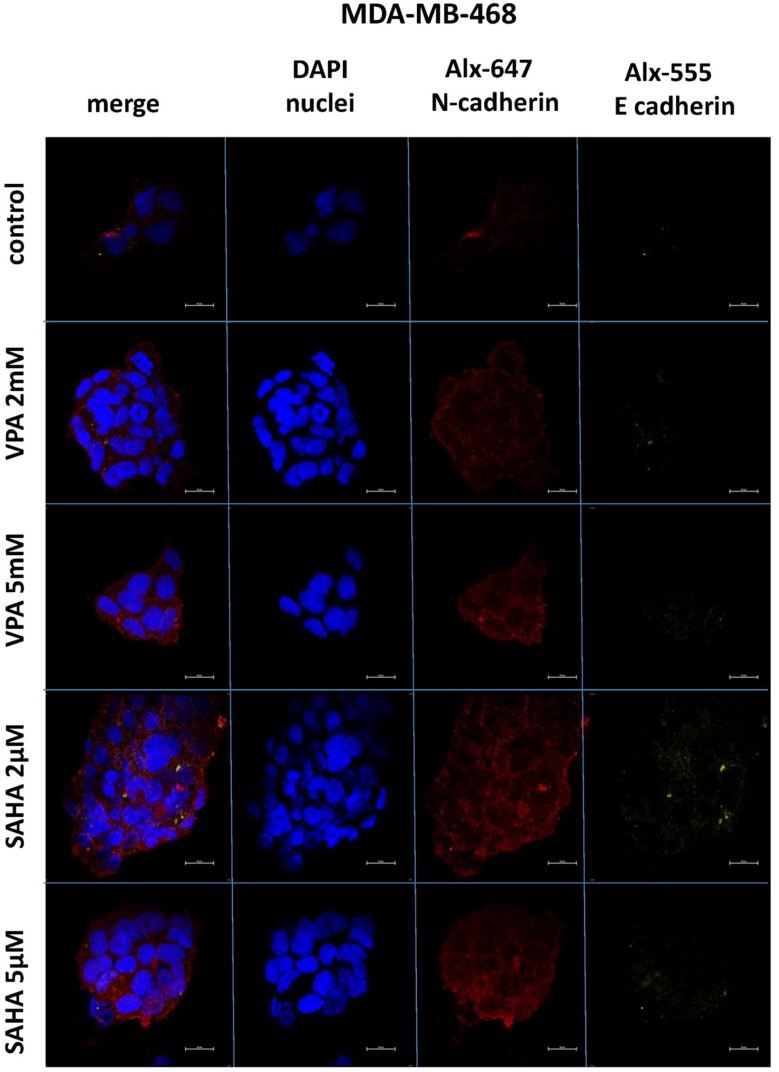
Figure 7.
Confocal imaging of E-cadherin and N-cadherin expression in MDA-MB-468 cells cultured alone (control) or in the presence of VPA (2 mM, 5 mM) and SAHA (2 µM, 5 µM).
Notes: Fixed cells were blocked and incubated with an anti-E-cadherin and anti-N-cadherin antibodies, followed by the respective secondary antibodies conjugated with Alexa fluorochromes as described in “Material and methods“ section. Nuclei were stained with DAPI. 100 x oil immersion objective was used. Representative images are shown.
Abbreviations: SAHA, vorinostat; VPA, valproic acid.

