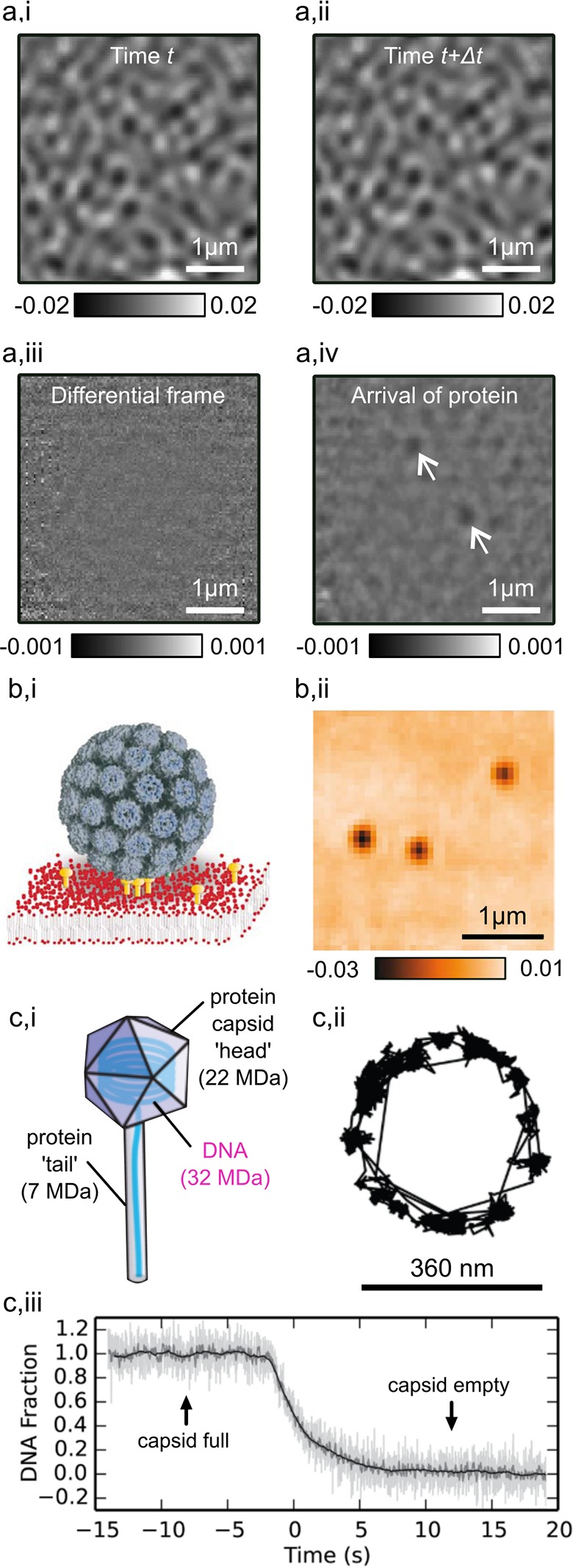
Figure 2.

iSCAT image of a bare coverslip recorded at a time t (a,i) and of the same area recorded at a later time t + Δt (a,ii). Subtraction of the image acquired at time t + Δt from the earlier frame removes static background features (a,iii). Dynamic arrival of two proteins registers in the differential frame (a,iv, marked with arrows). Adapted from ref (21). Illustration of a SV40 virus bound to ganglioside (GM1)-tagged lipids in an artificial lipid bilayer (b,i). An iSCAT image of single SV40 virions attached to a lipid bilayer on a coverslip (b,ii), adapted from ref (22). Illustration of a bacteriophage showing head and tail geometry (c,i). A trajectory from the head of a single bacteriophage whereas its tail is adsorbed to the surface (c,ii). Following stimulation, the DNA content of the capsid head is ejected over time (c,iii), as determined through the diminishing iSCAT contrast. Adapted from ref (44).
