
Figure 3.
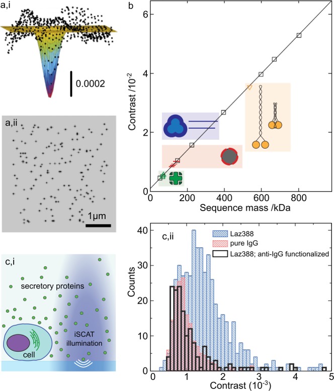
Sensitive detection of single proteins. (a,i) Nanometer localization of the PSF from a single protein. (a,ii) The super-resolved binding sites for many detected albumins across an imaging sequence. Adapted from ref (21). (b) Linear relation between the iSCAT signal and protein mass enables precise molecular weight calibration for different proteins and their complexes. Adapted from ref (25). (c,i) Illustration of the experimental arrangement for detecting the secretome from a single Laz388 cell. (c,ii) Histogram of contrast (mass) of secreted proteins, wherein the Immunoglobulin G (IgG) fraction can be identified. Adapted from ref (48).