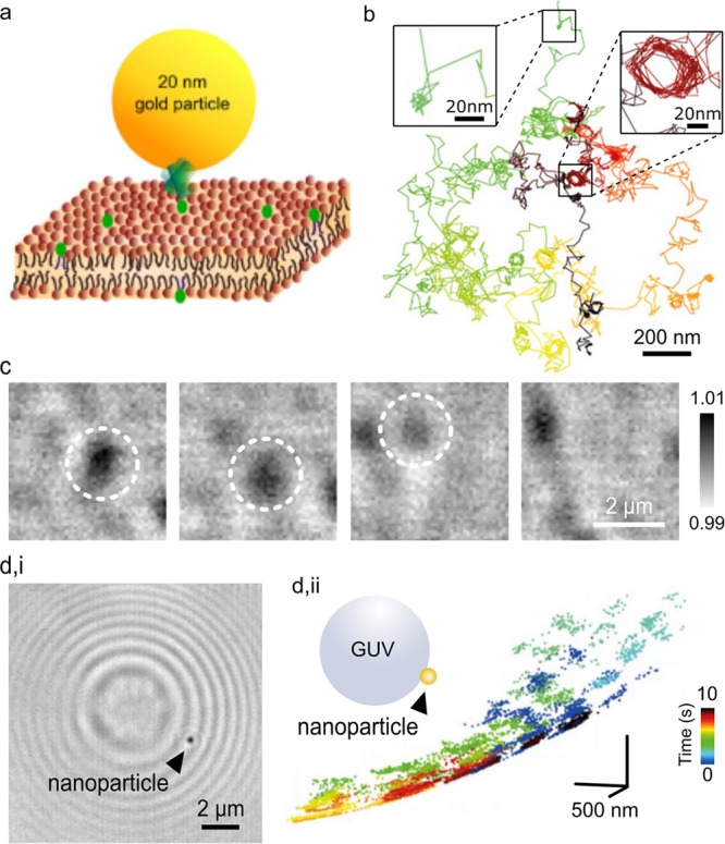
Figure 4.

iSCAT study of lipid bilayers. (a) Schematic of a single GNP labeling lipids via a linker in a lipid bilayer membrane. Adapted from ref (40). (b) Diffusive trajectory of a membrane lipid over 5 s recorded at 1000 fps, revealing examples of confinement into nanoscale geometries (insets). Adapted from ref (18). (c) Image sequence of a transient liquid-ordered nanodomain (marked with a dashed circle) disappearing into the surrounding lipid bilayer membrane, taken from ref (51). (d,i) Newton rings appear in a wide-field iSCAT image of a GUV. A single Tat-coated polymer nanoparticle (marked by arrow) is bound to the outer surface. (d,ii) Schematic of a nanoparticle attached to a GUV, and a three-dimensional diffusional trajectory of the Tat-coated polymer nanoparticle, showing Brownian diffusion on a spherical surface. Color coding indicates temporal progression. Adapted from ref (18).
