
Figure 6. P16 and P30 intractions close to the vertex.
(A) Shows the difference in interaction (pointed by red arrow) of the C-terminal Gly84 of P30 (orange) with P5 (bright pink), P31 (yellow) and P3 (green). The hydrophobic intraction of P30 with P5 is more obvious. (B) Shows the P3-P30-P16 complex (the view is similar to (A) but one copy of P16 and P30 are shown and the penton proteins are hidden), P16 (blue) locks the two adjacent P3 trimers (trimer 1)(green) and the P30 protein (orange).
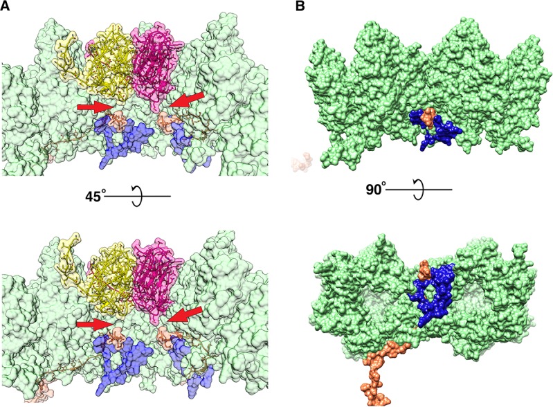
Figure 6—figure supplement 1. Partial model of P16 and its map density fit.

(A) The model of P16. (B) The stereo images of the model fitted into the CryoEM map.
Figure 6—figure supplement 2. P30 network.

(A) P30 monomer and its fit into the CryoEM density map. (B) interlocked N-terminal hook forming the dimer that spans between two adjacent vertices. Residues Met1-Val32 are involved the formation of the N-terminal hook. (C) Complete P30 cage that stabilizes the trisymmetrons and in turn the whole particle.
Figure 6—figure supplement 3. Difference density map showing the disordered region under the penton.

(A–C) Difference density map (blue) at the vertex region where the densities of the penton (P31 and P5) are shown in transparent gray. (A, D), (B, E) and (C, F) show the top, side and the bottom view of the difference density map, respectively.
Figure 6—figure supplement 4. Schematic of the infection mechanism model of bacteriophage PR772.

(A) Shows bacteriophage PR772 approaching the host membrane with the receptor. (B) The trimeric knob domain of P5 recognizes the host. The binding of the trimeric knob of P5 is transient. (C) High-affinity P2 binding to the surface receptor stabilizes the host binding. The binding is irreversible and attaches the virus to the host membrane. (D) P2 binding disturbs of the vertex complex by pulling the P5. (E) The disordered vertex complex disrupts the interactions between P5 and P30 resulting in a cascade that leads to the disruption of the P30 cage that stabilizes the viral capsid. (F) Disrupted P30 cage, destabilizes other adjacent vertices and the interactions that anchor the viral membrane. (G) P16 anchors into the host membrane and facilitates the formation of the membranous tube. (H) Destabilized viral membrane collapses and the dsDNA is delivered into the host.
Figure 6—video 1. Movie showing the P3-P30-P16 complex.
Download video file (8.4MB, mp4)
DOI: 10.7554/eLife.48496.028