Figure 4.

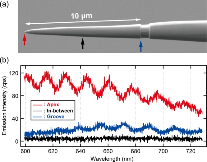
STL spectra recorded at different positions on the grooved tip. (a) SEM image of the grooved tip with L = 10 μm. (b) STML spectra obtained at the apex (red), the groove (blue), and in-between (black) (Vbias = 2.5 V, It = 9 nA).

STL spectra recorded at different positions on the grooved tip. (a) SEM image of the grooved tip with L = 10 μm. (b) STML spectra obtained at the apex (red), the groove (blue), and in-between (black) (Vbias = 2.5 V, It = 9 nA).