
Fig. 5. FXR1 overrides CDK4/6i-induced senescence through SLC36A1.
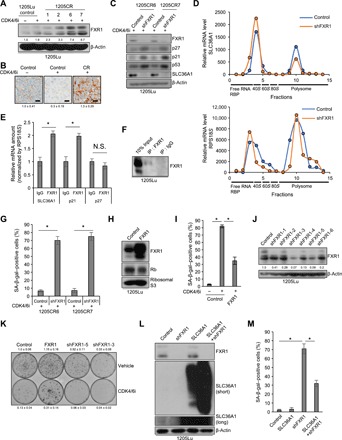
(A) Western blot analysis of lysates from 1205Lu cells treated with or without palbociclib (1 μM) for 8 days and 1205CR1, 1205CR2, 1205CR6, and 1205CR7 cells using antibodies to FXR1 and β-actin. The numbers indicate quantifications of FXR1 determined by FXR1/β-actin ratio. (B) Representative images of IHC staining of sections from xenograft tumors treated with either vehicle or palbociclib (1 μM) or tumors resistance to CDK4/6i for FXR1. Scale bars, 50 μm. The numbers indicate quantification of FXR1 intensity determined by IHC scoring (see Materials and Methods) from three independent experiments. (C) Western blot analysis of lysates from 1205CR6-7 cells with or without shFXR1 using antibodies indicated on the right of the panels. (D) Fractions from HEK293T cells with or without shFXR1 were collected by density gradient fractionation. Purified RNA was subjected to qRT-PCR analysis using sets of primers for SLC36A1 (top) and RPS18S (bottom). This experiment was performed three times, and representative data were shown. RBP, ribose-binding protein. (E) Lysates of 1205Lu cells were analyzed by RIP assay using antibodies to control IgG or FXR1 and subjected to qRT-PCR using sets of primers for SLC36A1, p21, and p27. Data were normalized by IgG and represent means ± SD, *P < 0.01 (one-sample two-tailed Student’s t test; n = 3). (F) Western blot analysis of the lysates from (E) using antibodies to FXR1. (G) Quantification of SA-β-gal–positive cells in 1205CR6-7 cells introduced with shcontrol (control) or shFXR1. Data represent means ± SD, *P < 0.01 (two-tailed Student’s t test; n = 3). (H) Western blot analysis of lysates from 1205Lu cells introduced with empty vector (control) or FXR1 using antibodies to FXR1, Rb, and ribosomal S3. (I) Quantification of SA-β-gal–positive cells in 1205Lu cells introduced with vehicle (control) or FXR1 with or without palbociclib treatment (1 μM) for 8 days. Data represent means ± SD, *P < 0.01 (two-tailed Student’s t test; n = 3). (J) Western blot analysis of lysates from 1205Lu cells with shcontrol or shFXR1 using antibodies to FXR1 and β-actin. The numbers indicate quantifications of FXR1 determined by FXR1/β-actin ratio. (K) Clonogenic colony formation assay of cells from (H) and (J) treated with or without palbociclib (1 μM) for 8 days. The numbers indicate quantification of colonies from three independent experiments. (L) Western blot analysis of lysates from 1205Lu cells infected with shcontrol, shFXR1, SLC36A1, or shFXR1 + SLC36A1 using antibodies to FXR1, SLC36A1, and β-actin. (M) Quantification of SA-β-gal–positive cells from (L). Data represent means ± SD, *P < 0.01 (two-tailed Student’s t test; n = 3).